Wamkelekile, Welkom
Inside growing brains
22 November 2019 | Story Ambre Nicolson. Photo Tina Floersch, Unsplash. Read time 4 min.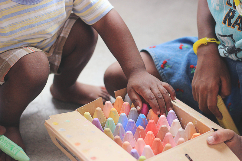
Recent growth in availability of safe and non-invasive techniques for visualising the brain has had a huge impact on how we study children’s brains. UCT researchers Professor Kirsty Donald and Professor Ernesta Meintjes explain what they have learnt about how young brains develop in high-risk contexts.
“In the past, it wouldn’t have been ethically responsible to expose children to even the small amount of radiation that CT scanners emit unless there was a clinical indication to do so,” explains Donald, a paediatric neurologist based at the Red Cross War Memorial Children’s Hospital in Cape Town. “But magnetic resonance imaging (MRI) has made it possible for us to conduct this kind of research.”
“One major benefit of MRI – which is completely safe, non-invasive and does not involve any harmful, ionising radiation – is that we can ethically study brain development and ageing in normal, healthy populations from birth, throughout childhood, into adulthood and beyond,” agrees Meintjes, the South African Department of Science and Technology/National Research Foundation Research Chair in Brain Imaging.
“This allows us to establish trajectories of normal brain development and ageing – critical if we want to examine how disease alters brain development and function.”
“Magnetic resonance imaging has made it possible for us to conduct this kind of research.”
Donald and Meintjes are involved in several long-term studies looking at how children’s brains develop – both in the womb and after birth – in the context of high-risk factors like alcohol use, cigarette smoking, methamphetamine use, poor maternal mental health and exposure to HIV.
For example, they have shown that exposure to HIV can alter the health and maturity of white matter – which affects learning and brain functions – in babies’ brains, even if they have not been infected by the virus.
“It looks like this is also leading to structural changes later on,” says Donald.
Meintjes and Donald have also looked at how alcohol affects the brain in utero – when it is at its most plastic. They have imaged the brains of children from a very early age to understand how changes resulting from exposure to alcohol come about and progress to differences among school-aged children.
Meintjes explains that recent research from the Cape Universities Body Imaging Centre, of which she is director, showed that particular supplements can help mitigate the adverse effects of alcohol exposure for babies whose mothers consumed large amounts of alcohol while they were pregnant. These babies grew better and performed better cognitively if their mothers took supplements of choline – a nutrient found in many foods.
Both of these studies were made possible by neuroimaging techniques.
“Neuroimaging allows us to investigate relationships between imaging measures, such as brain volumes, with neuropsychological performance, neurocognitive function, socioeconomic measures, inflammatory and genetic markers, microbiome diversity, et cetera,” continues Meintjes.
“In the future, this will help us to figure out the brain activity related to specific diseases and disorders.”
 This story was published in the fourth issue of Umthombo, a magazine featuring research stories from across the University of Cape Town.
 Umthombo is the isiXhosa word for a natural spring of water or fountain. The most notable features of a fountain are its natural occurrence and limitlessness. Umthombo as a name positions the University of Cape Town, and this publication in particular, as a non-depletable well of knowledge.
Read the complete fourth issue online or subscribe and receive new issues in your inbox every few months.
 This work is licensed under a Creative Commons Attribution-NoDerivatives 4.0 International License.
Please view the republishing articles page for more information.


Research & innovation