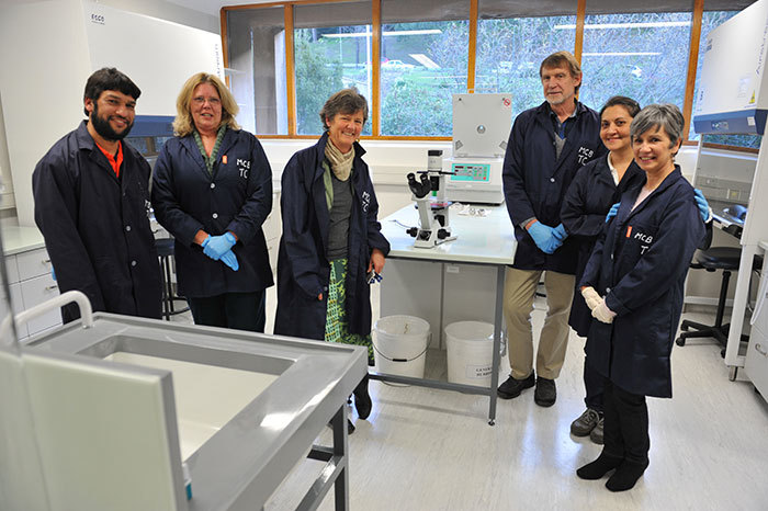
Renovated laboratory boasts world-class facilities and equipment
20 October 2014 | Story by Newsroom
An upgraded mammalian tissue facility with state-of-the-art equipment recently opened at UCT. Mammalian tissue culture involves experiments with either primary animal cells or cell lines that require sterile conditions, to not only protect the experimental reagents from contamination, but also to safeguard the scientist from exposure to potentially bio-hazardous agents.
"These studies require specialised equipment within a dedicated facility restricted to specific experiments under the guidance of stringent internationally recognised health and safety regulations," says Professor Janet Hapgood of the Department of Molecular and Cell Biology (MCB) (MCB), whose research was one of the drivers that led to the renovation of the facility.
Established in the late 1970s, the old facility could not support the growing interest amongst postgraduates wanting to carry out research in human health, especially those focused on infectious diseases. The entire facility was reduced to a shell before it was upgraded to international standards, a process that included the restructuring, expansion and upgrading of the existing air-conditioning system.
The upgrade of the facility was made possible through the efforts of three key players: architect and project manager Gloria Robinson, principal scientific officer Faezah Davids, and principal technical officer Neil Bredekamp. They worked tirelessly to secure the professional expertise required to design and configure the new facility. Without the contribution of the dynamic and innovative MCB Life Sciences Workshop team the facility would also not have been possible, adds Hapgood.
"The facility has greatly increased our competitive edge in the field of basic research in HIV-1 pathogenesis and human papillomavirus and other vaccinology, access to international funding, recruitment of students, and has added to the undergraduate curriculum. We are very proud of it, and are grateful to all those involved in funding, building and enabling it," says Hapgood.
Cutting-edge equipment
An inverted fluorescent microscope, housed in a dedicated microscope room in the facility, is an example of the state-of-the-art equipment now available. Paired with a mini-incubator, this microscope enables researchers to directly visualise live mammalian cells, and is set to become an invaluable resource to researchers at UCT. The microscope can obtain high-contrast and high-resolution images at a range of magnifications, as well as narrow band filter blocks to allow fluorescent imaging in the ultraviolet, fluorescent green and red range.
Images captured by the microscope on one of three cameras are then stored on a dedicated 10-terabyte server named ROSALIND, housed in the UCT data centre, and named in memory of Rosalind Franklin who captured the first X-ray diffraction images of the DNA double helix.
All data stored on this dedicated server is then automatically backed up for six weeks from the date of acquisition. There are two options to view imaging data on the ROSALIND Date Store. Users can download the open source OMERO software to view and manage their image data, or access NIS-Elements imaging software via a licencing dongle. Not only does this give researchers the flexibility of using this sophisticated software from their workstations in their laboratories, a safe storage solution has also been found for this expensive dongle in the ICTS data centre.
Image by Michael Hammond.
 This work is licensed under a Creative Commons Attribution-NoDerivatives 4.0 International License.
Please view the republishing articles page for more information.


