5 questions with Tinashe Mutsvangwa, biomedical engineer
14 February 2020 | Story Lisa Boonzaier. Photo Brenton Geach. Read time 6 min.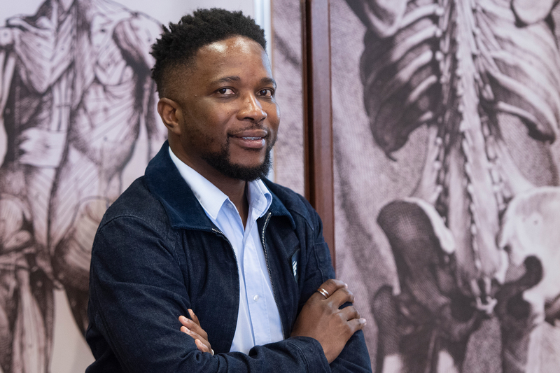
As group lead for Medical Image-based Inferencing and Distributed Diagnosis in the University of Cape Town’s (UCT) Division of Biomedical Engineering, Dr Tinashe Mutsvangwa has established himself as an expert in the development of advanced medical imaging and analysis. He was recently elected to the highest professional membership grade of the Institute of Electrical and Electronics Engineers.
1. What does the field of medical imaging and analysis encompass?
Medical imaging encompasses the use of various technologies (including X-ray, ultrasound and even mobile phones) to view the human body for aid in diagnosis, monitoring and treatment of medical conditions. For the analysis component, we develop computational algorithms to manipulate the images with the aim of enhancing and presenting the most relevant information that can assist clinicians to make the best decisions for their patients.
2. What impact does your research have on the everyday lives of people?
My colleague, Professor Tania Douglas, and I are working on two major medical imaging applications. One is the 3D reconstruction of computed tomography (CT) images from regular 2D X-rays. The other is mobile phone-based testing for latent tuberculosis. Once matured, we envision the first will lead to better access to advanced medical imaging – at a lower cost. The second aims to improve screening for latent tuberculosis (TB).
The diversity of experience among collaborators always brings depth to research.
3. What is the biggest opportunity you see for your field?
Globally, well-resourced countries have ready access to state-of-the-art medical-imaging resources, while people living in low- to medium-income countries have much less access to basic medical imaging. I see an opportunity to bridge this divide by leveraging machine learning and the ever-growing archives of medical images from different imaging technologies.
One of our main research thrusts is to use algorithms to synthesise images of one type of imaging technology from another. For example, synthesising a more expensive modality from a cheaper one that’s more available – like CT from 2D X-ray, as I mentioned above. This would be very useful for under-resourced healthcare systems.
4. What is the most enjoyable aspect of your research?
A significant part of medical image analysis involves developing computer algorithms to automate the identification and quantification of pathology in images. The most enjoyable part is brainstorming with my students on my whiteboard to come up with novel algorithms. It is extremely gratifying when mathematical ideas (expressed as computer code) produce the desired results in terms of quantifying or classifying a medical condition from medical images.
5. What advice do you have for young researchers?
Find a good network of collaborators from around the world in your research area. Collaborators provide an opportunity for a wider pool of resources, whether its grants, equipment or software. The diversity of experience among collaborators always brings depth to research. And collaborators provide opportunities for exchange, which makes your research group more attractive to students and provides you with opportunities to travel.For me, this Sutherland project was an example of engaged scholarship of the highest pedigree; a multi-disciplinary team bringing their expertise to bear on an initiative of significance cultural importance. The values I was referring to were justice for the descendants of the individuals, community-centredness on the part of the scientists in terms of how they approached the project and accountability on the part of UCT accepting the historical role it played in the unethical acquirement of the remains.
Read more about Mutsvangwa’s involvement in this project.
 This work is licensed under a Creative Commons Attribution-NoDerivatives 4.0 International License.
Please view the republishing articles page for more information.


