Wamkelekile, Welkom
Seeing the brain through the eyes
26 November 2018 | Story Nadia Krige. Photo Pexels, Pixabay. Read time 10 min.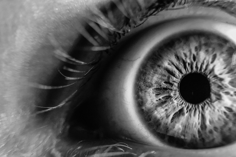
Since ancient times, the eye has been seen as a portal to the innermost secrets of the human mind and body – a window to the soul. Taking a more literal approach to this metaphorical truth, Dr Llewellyn Padayachy, a paediatric neurosurgeon at UCT, has spent the past five years or so researching a novel diagnostic method that uses ultrasound and the eye as a window to the brain.
During the most recent instalment of Café Scientifique – hosted by UCT Research Contracts and Innovation (RC&I) and supported by leading intellectual property law firm Spoor & Fisher – Padayachy introduced his findings and shared some insights into his invention journey.
“What you do as a paediatric neurosurgeon is treat a condition called hydrocephalus more than anything else,” Padayachy explained. “And the sad thing is, a lot of the children we treat, without a doubt, present to us much later than they should.”
More often than not, the reason for this is that the children come from impoverished rural areas, where the condition would not have been picked up in a primary healthcare clinic, due to a lack of resources.
While accurate data regarding the number of hydrocephalus cases on the African continent is sparse, conservative estimates put it at some 100 000 cases annually.
While accurate data regarding the number of hydrocephalus cases on the African continent is sparse, conservative estimates put it at some 100 000 cases annually. In the United States, it affects about one million people in every stage of life: from infants to the elderly.
One of the first indications of hydrocephalus in children is raised pressure inside their skulls. And up until recently, the gold standard for measuring this has been drilling a hole in the skull, placing a catheter in the brain and directly measuring the pressure.
Undergoing this sort of procedure is highly traumatic for any patient and even more so for babies and young children.
Witnessing this procedure first-hand on a daily basis inspired Padayachy to set out in search of a non-invasive means to measure pressure in the brain. Furthermore, he wanted it to be reproduceable and simple enough to use at primary healthcare clinics in rural settings to help diagnose hydrocephalus earlier.
Journey of discovery
With its connection to the brain via the optic nerve, the eye can serve as a gateway for physicians and surgeons.
Dr Llewellyn Padayachy, a paediatric neurosurgeon at UCT, has spent the past five years or so researching a novel diagnostic method that uses ultrasound and the eye as a window to the brain
Unsurprisingly, Padayachy’s journey toward developing a ‘holy grail’, non-invasive technique to assess pressure inside the skull started with the eye.
At the time, he had been using ultrasound as an intraoperative guidance tool to improve surgical precision – both for tumours and hydrocephalus. This led him to explore its further diagnostic potential as a non-invasive tool.
So Padayachy and team, which include Norwegian engineers Tormod Selbekk and Reidar Brekken, who were introduced to each other many years ago by UCT Professor Graham Fieggen, started using a small ultrasound probe to scan about 2.5 centimetres behind the eye, allowing them to view the optic nerve and the cerebrospinal fluid that surrounds it – and analyse this dynamic relationship as a marker for brain pressure.
“Raised intracranial pressure is effectively a progressive build-up of pressure in the brain to the point where the normal brain, fluid and blood volume relationship is displaced in some sort of proportion,” Padayachy explained. “The first compartment to be displaced is cerebrospinal fluid.”
Witnessing this procedure first-hand on a daily basis inspired Padayachy to set out in search of a non-invasive means to measure pressure in the brain.
The eye is directly linked to the brain via the optic nerve, which sits at the back of the eyeball. As pressure in the brain builds up, cerebrospinal fluid is forced along the optic nerve sheath, which then dilates in the same way as a balloon inflates.
“We figured that if this sheath appeared ‘stretched’ when we compared it to other clinical and imaging markers, then this information could be really useful.”
During an inspiring year at Oxford University, as part of his Hamilton Naki Clinical Scholarship in 2016/2017, Padayachy studied the use of magnetic resonance imaging and other novel, non-invasive techniques using the visual pathway to assess brain function. Thereafter, he returned to Cape Town to round up some of his work with researchers at SINTEF, an independent research organisation in Trondheim, Norway.
In addition to static images, the team started collecting 10-second video clips to capture the pulse-related motion of the nerve sheath behind the eye. In combination, these static and dynamic images would allow them to measure the diameter of the nerve sheath as well as its stiffness, significantly advancing current thinking in this field.
“If you have a sheath that is wide and stiff, it is more predictive of raised intracranial pressure than a sheath that is wide and quite [relaxed],” explains Padayachy.
... it turns out that this tool could be useful in a surprising number of other settings.
This formula for defining intracranial pressure is central to the team’s patent, which takes the form of a software package that enables users to harness ultrasound data efficiently and effectively. This has not only lead to Padayachy’s PhD and a number of publications, but also to the creation of spinoff company NISONIC (Non-Invasive SONographic IntraCanial pressure monitor).
The team’s commercialisation company is currently in the process of acquiring the European Conformity mark, after which they aim to get prototypes equipped with the patented software available in the next two years.
From rural clinics to the final frontier
While Padayachy’s invention was inspired by the dream of offering primary healthcare clinics in rural settings the opportunity to diagnose raised intracranial pressure and possible hydrocephalus in children early on, it turns out that this tool could be useful in a surprising number of other settings.
NASA has recently found that astronauts in space develop visual impairment due to raised intracranial pressure, which can prove to be life-threatening in a job where being able to see is clearly of the utmost importance.
Thus, having a non-invasive, portable tool that can measure intracranial pressure in space would – no doubt – be hugely useful.
“If I could indulge a pipe dream, it would be that we have a little South African imprint on the first mission to Mars where we use this method of diagnosing raised intracranial pressure,” says Padayachy. “It would put a feather in the cap of UCT, Cape Town and South Africa.”
 This work is licensed under a Creative Commons Attribution-NoDerivatives 4.0 International License.
Please view the republishing articles page for more information.