Wamkelekile, Welkom
Mammo-graphy goes digital
19 April 2005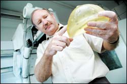
Novel technology: Prof Kit Vaughan with a breast phantom used to test the efficacy of mammography systems. The darker spots in the gel are simulated lesions representing cancers-related pathology of various sizes and densities. It is hoped that the new digital mammography techniques will detect smaller lesions, identifying cancers earlier. Using the circular scanning techniques that Vaughan's team is working on will hopefully also make the procedure far more comfortable for patients.
Women who have undergone routine mammograms via conventional screen-film methods will know that the procedure is not only uncomfortable but often painful. The cancerous lesions can also sometimes escape detection. But a group of UCT researchers, working with the private sector, hopes to change that - by covering a wider area of the breast and bumping up the quality of X-ray images using novel slot-scanning techniques.
It is also hoped that these new scanning techniques could reduce the need for breast compression, normally necessary to minimise scattered radiation (photons that muddy the X-ray picture, producing what radiographers refer to as "noise"), as well as reduce the thickness of the breast and immobilise it.
The funder of the two-phase project of approximately
Innovations in the field are crucial. With significant intellectual property spin-offs (held by African Medical Imaging, of which UCT is a 44% shareholder) the new technology lists other potential advantages: lower radiation dose for patients and health-care staff, greater coverage of the breast, improved spatial resolution, reduced patient examination time, and an opportunity for computer-assisted diagnosis.
The technology is novel, developed by postgraduate students Mark Seymour and Mattieu de Villers, with each holding an international patent and others in the offing.
"Breast cancer accounts for 9% of cancers worldwide, and although recent media reports have questioned the benefits of mammographic screening, the scientific literature is clear: early detection and treatment of breast cancer have cut mortality rates by 20% in the past decade," notes principal investigator Professor Kit Vaughan (human biology).
Although a Cancer Association of South Africa report in 2000 reported that carcinoma of the breast was the commonest cancer in South African women, no national screening programme exists. Vaughan cites US figures of carcinoma of the breast resulting in 18% of cancer deaths. "This translates into 40 000 US women dying from the disease each year."
Digital mammography is not new - US researchers planted their flag on this territory some time ago with the US Food and Drug Administration's approval of the first "full-field" digital scanner in 2000. But it is recent enough to have quickened the imaginations of the UCT-led interfaculty team, keen to explore new technological refinements.
The MRC/UCT Medical Imaging Research Unit also has a reputation for innovation. With another five-year season of MRC funding, they have the scope to produce more of the original work that won Vaughan and Professor Gerhard de Jager (electrical engineering) the National Science and Technology Forum's trophy for innovations in medical imaging in 2002.
Phase 1 of the NIH-funded project will see the construction, design and testing of a prototype digital mammography system, one that will reduce radiation exposure while maintaining image quality, this by incorporating a patented slot-scanning technique recently approved for whole-body trauma applications by the FDA.
"We will incorporate solid-state image sensors in the design of the X-ray detectors, maximizing the signal-to-noise ratio," Vaughan explains.
Breast phantom studies and X-ray dose measurements will allow the performance of the digital mammography system to be optimised for the detection of small lesions.
"By scanning in a motion that accounts for breast anatomy, it will hopefully be possible to improve patient comfort by reducing the need for breast compression and also by covering a greater volume of breast tissue."
Because the images are digital, this will facilitate computer-aided diagnosis (CAD) while also reducing patient examination time.
The project is also about partnerships, as the multi-faceted group tackles the project from all angles. As co-investigators, commercial partner Lodox Systems (Pty) Ltd, represented by Bill Greenway and Herman Potgieter, contributes both manpower and creative ideas. Greenway, CEO of Lodox Systems, is a pioneer in digital mammography in the US, while Potgieter currently serves as honorary professor in UCT's Department of Electrical Engineering.
Another key person is Dijon-born researcher and PhD in experimental particle physics from the University of Glasgow, Dr Julien Marchal. Though the postdoctoral fellow talks admiringly of new theory development at CERN in Geneva and the Fermi National Accelerator Laboratory in the States, it's the human applications of radiation detection that motivate him.
In Glasgow, for example, Marchal contributed to a project to create artificial retinas using pixel detectors, involving microchip implants in the retina.
Writing in Elektron (March, 2005), Marchal explained: "The tight requirements in terms of image quality and X-ray dose have made mammography one of the last medical X-ray imaging examinations to date to fall under the pressure of the 'digital revolution'."
Marchal will explore the full potential of digital techniques in mammography. A novel scanning configuration, invented by Seymour, will provide greater coverage of beast anatomy. This circular scanning allows a larger picture of breast tissue, for example, towards the region of the armpit, where cancerous nodes often form and which conventional techniques may miss, all at superior resolution levels.
"With a digital system you can really manipulate the image, enlarging it, adjusting the grey scale to enhance the clarity of subtle structures in the breast."
He adds: "We're developing new ways of scanning the breast that follow the shape of the chest wall." And good news for patients and health care workers is that the digital system provides a lower dose of X-rays compared to conventional methods.
During the last six months of the project in phase 2, the group will conduct a limited clinical trial of 20 patients, focusing on comfort, breast coverage and diagnostic equivalence.
"The long-term aim of this project is to address the drawbacks of current screen-film mammography devices by developing a digital mammography system based on innovative medical imaging technologies arising in South Africa," Vaughan explains.
Dr Tania Douglas (human biology), an expert in medical imaging and co-investigator, agrees.
"The idea behind the project is that we are confident we can improve on existing systems."
Professor Steve Beningfield, head of radiology at Groote Schuur Hospital, and also a co-investigator on the grant, says: "The technology offers interesting and unique methods of overcoming some of the important limitations of film mammography, while promising top-quality, large field-of-view digital images."
The results will hopefully signal a transfer to a more effective, modern and patient-friendly imaging tool.
(Additional information source: http://imaginis.com/breasthealth/digital_mammo.asp)
 This work is licensed under a Creative Commons Attribution-NoDerivatives 4.0 International License.
Please view the republishing articles page for more information.


