Wamkelekile, Welkom
New hermit crab species in 3D
29 October 2018 | Story Helen Swingler. Read time 6 min.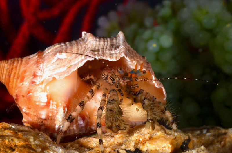
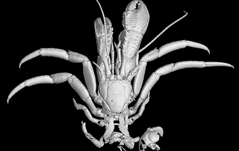
Three-dimensional X-ray micro-computed tomography has been used to describe a new species of hermit crab, Pagurus fraserorum, found in the rocky subtidal reefs off the coast of KwaZulu-Natal (KZN).
The study, by University of Cape Town (UCT) marine taxonomist Dr Jannes Landschoff (Department of Biological Sciences), is the first description of a hermit crab in which most of the taxonomic details have been illustrated using 3D volume-rendered illustrations: added line drawings to show high-resolution surface structures.
The paper was published in PLOS One. Landschoff’s co-authors are UCT colleague Emeritus Professor Charles Griffiths; Tomoyuki Komai of the Natural History Museum and Institute in Japan; Anton du Plessis of the CT Scanner, Central Analytical Facility, Stellenbosch University; and Gavin Gouws of the National Research Foundation – South African Institute for Aquatic Biodiversity.
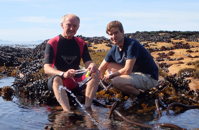
The new crab species has been named after Mike and Valda Fraser and their son Allan, well-known in the dive community and authors of several reef guides. The Frasers hosted Landschoff and Griffiths during their collection trip in October 2015, when the species was first recorded.
“The naming honours their deep passion for the marine world, and the many exciting species discoveries they’ve made in South African waters,” said Landschoff.
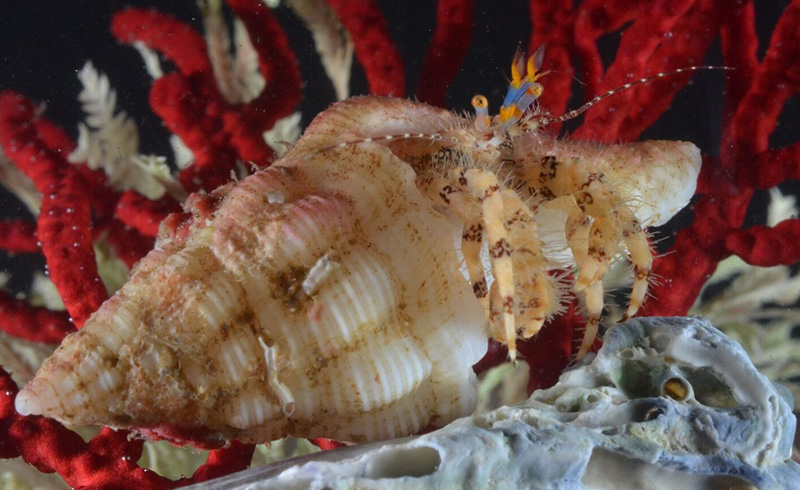
Landschoff has now described three new species of hermit crab, including a green-eyed deep-water hermit crab, Paragiopagurus atkinsonae, found only in a tiny area off the West Coast of South Africa.
Relatives in Indo-Pacific
The new species, pinkish-maroon and cream in colour, was found among the rocky reefs off Pumula and Hibberdene near Port Shepstone in KZN. It was known previously only from a photographic record taken at Veitch’s Pier in Durban.
It is the sixth species of Pagurus known in South Africa and is closely related to (but genetically distinct from) the Indo-Pacific crabs Pagurus boriaustraliensis, from north and north-western Australia, and specimens of Pagurus pitagsaleei, from Taiwan.
However, with its characteristic shape, spines and hairs on the pincers and the walking legs, Pagurus fraserorum in South Africa is not easily mistaken for any other, said Landschoff.
X-ray micro-computed tomography, or Micro-CT, has become one of the most powerful tools for generating 3D illustrations. It’s been useful in morphological studies of various other invertebrate groups such as insects and spiders, and has also been applied to marine invertebrates such as sponges, bivalves (aquatic molluscs with hinged shells, such as oysters) and sea urchins.
The technique gives taxonomists a new tool to “dissect” and study rare or delicate specimens on their computer screens and with or without museum or natural history collections and artists’ replicas.
It won’t fully replace physical specimens, but it does allow scientists to download interactive, printable scans – or digital avatars – for 3D examination of specimens that can be studied simultaneously by people in different places. Also, actual specimens – many rare and valuable – are difficult to transport.
Soft tissue issue
Ironically, hermit crabs are among the most difficult model organisms within the higher crabs on which to use CT scanning. Many are small, and half of the body – the pleon or tail, which is encased in the scavenged sea shell it lives in – is soft, so the structures are difficult to detect.
The authors write that in higher taxa of crustacea, the techniques have been used to study sexual reproductive organs or internal anatomy, such as the complex lung system of the terrestrial fiddler crab and the vascular system and inner anatomy of Anomura, which includes hermit crabs.
Not all taxonomic characters can be properly shown using 3D. In this case, however, overlaid manual drawings were a viable solution, combining the benefits of both the historical and modern taxonomic techniques.
“The 3D data is thus referred to as 3D type data, to be clear that they are derived from the physical specimen,” Landschoff explained.
Composite visuals
They showed, he added, that hermit crabs, and certainly most other crustacea, are in fact suitable candidates for this now well-established method to be applied in taxonomy.
“Whether one of the main benefits of 3D Micro-CT – the sharing of 3D data of valuable specimens – will advance and streamline information exchange, and to some extent replace the difficult process of loaning physical specimens, must be explored further.
“This might happen only when most taxonomic experts have access to scanning facilities, as well as to the necessary software for processing 3D Micro-CT data.”
 This work is licensed under a Creative Commons Attribution-NoDerivatives 4.0 International License.
Please view the republishing articles page for more information.


Research & innovation